lh3.googleusercontent.com
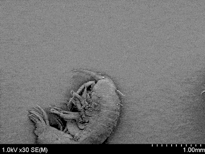
A bacterium on a diatom on an amphipod
Scanning electron microscopy (SEM) is a powerful imaging technique that produces images of a sample by scanning it with a beam of electrons instead of light. The electrons interact with the atoms in the sample to produce signals containing information about the sample’s surface topology and composition. Because electrons are much smaller than the wavelength of visible light, SEM can achieve resolutions better than 1 nanometer (100,000x thinner than human hair).
Here, an SEM is used to zoom in on a prepared sample by several orders of magnitude. The microscope initially reveals an amphipod, a type of shell-less crustacean (1mm). As it zooms in on the head of the amphipod, a round, silica-shelled algae called a diatom is revealed (100µm). As it zooms in even further, a rod-shaped bacterium can be seen resting on the diatom (500nm).